The human body is an unparalleled masterpiece of biological engineering, a complex network of systems working in perfect synchrony to maintain life. From the microscopic filtration units in the kidneys to the vast neural highways of the brain, every organ plays a specific and vital role. Understanding these components is not merely a matter of medical interest but a fundamental requirement for maintaining long-term health and wellness. This pillar page serves as a comprehensive repository of knowledge regarding the most critical organs and tissues that constitute the human experience.
The Thyroid Gland: Master of Metabolic Regulation
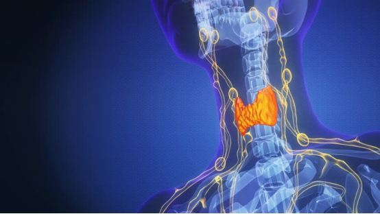
Located in the anterior neck, the thyroid gland is a butterfly-shaped endocrine organ that exerts profound influence over nearly every physiological process in the body. Its primary function is the production of hormones that regulate the rate at which the body uses energy, a process known as metabolism. By secreting thyroxine (T4) and triiodothyronine (T3), the thyroid dictates the speed of cellular activities, affecting body temperature, heart rate, and weight management.
Hormonal Synthesis and Iodine Uptake
The production of thyroid hormones is a multi-step process that requires iodine, a trace element obtained through diet. The gland traps iodide from the bloodstream and combines it with the amino acid tyrosine to create T3 and T4. This process is strictly regulated by the Thyroid-Stimulating Hormone (TSH) released by the pituitary gland. When thyroid hormone levels are low, TSH levels rise to stimulate production; conversely, high levels of thyroid hormones trigger a reduction in TSH secretion through a negative feedback loop.

Understanding Thyroid Disorders and Health
Thyroid dysfunction is common and can manifest in two primary forms: hypothyroidism and hyperthyroidism. These conditions represent the extremes of metabolic activity. Hypothyroidism occurs when the gland is underactive, leading to symptoms such as fatigue, weight gain, and depression. Hyperthyroidism, or an overactive thyroid, accelerates the body’s processes, causing rapid heartbeat, anxiety, and unintended weight loss.
Common Thyroid Pathologies
- Hashimoto’s Thyroiditis: An autoimmune disorder where the immune system attacks thyroid tissue, leading to chronic inflammation and hypothyroidism.
- Graves’ Disease: An immune system disorder that results in the overproduction of thyroid hormones, often characterized by bulging eyes (exophthalmos).
- Thyroid Nodules: Lumps that form within the gland; while most are benign, a small percentage can be cancerous and require surgical intervention.

The Pancreas: Dual Roles in Digestion and Endocrine Function
The pancreas is a unique organ situated behind the stomach that functions as both an exocrine and endocrine gland. Its exocrine tissues produce digestive enzymes that break down proteins, fats, and carbohydrates in the small intestine. Simultaneously, its endocrine clusters, known as the Islets of Langerhans, secrete hormones directly into the bloodstream to regulate glucose levels.
Exocrine Secretions and Enzyme Activity
The pancreas produces approximately 1.5 liters of pancreatic juice daily. This fluid contains bicarbonate to neutralize stomach acid and enzymes such as amylase (for starches), lipase (for fats), and proteases like trypsin and chymotrypsin (for proteins). Without these secretions, the body cannot effectively absorb nutrients, leading to malabsorption syndromes.

Pancreatic Enzymes and Insulin Management
The endocrine function of the pancreas is centered on blood sugar homeostasis. The two primary hormones involved are insulin and glucagon. Insulin is produced by beta cells and acts as a “key” that allows glucose to enter the body’s cells for energy use. Glucagon, produced by alpha cells, stimulates the liver to release stored glucose when blood sugar levels drop too low.
| Hormone | Cell Type | Primary Function |
|---|---|---|
| Insulin | Beta Cells | Lowers blood sugar by promoting glucose uptake. |
| Glucagon | Alpha Cells | Raises blood sugar by stimulating glycogenolysis. |
| Somatostatin | Delta Cells | Inhibits the release of both insulin and glucagon. |

The Spleen: The Body’s Blood Filter and Immune Guardian
The spleen is the largest organ in the lymphatic system, located in the upper left quadrant of the abdomen. Often overlooked, it serves as a critical quality control center for the blood. The spleen filters out old, damaged, or malformed red blood cells, ensuring that only healthy cells circulate through the cardiovascular system.
Red Pulp vs. White Pulp
The spleen’s tissue is divided into two distinct types: red pulp and white pulp. The red pulp is responsible for the mechanical filtration of blood and the storage of platelets and monocytes. The white pulp is composed of lymphatic tissue and is the site where immune responses are mounted. It contains B-cells and T-cells that identify and attack pathogens such as bacteria and viruses present in the bloodstream.

Functions of the Spleen in Lymphatic Health
Beyond filtration, the spleen acts as a reservoir for blood. In the event of a traumatic injury causing significant blood loss, the spleen can contract and release its stored supply of red blood cells and platelets into circulation. This “auto-transfusion” can be a life-saving mechanism during acute hemorrhagic shock.
The Impact of Splenectomy
While the spleen performs vital functions, it is not essential for life. If the spleen is removed (splenectomy) due to rupture or disease, other organs like the liver and lymph nodes take over most of its filtration duties. However, individuals without a spleen are at a significantly higher risk for certain bacterial infections, particularly those caused by encapsulated organisms like Streptococcus pneumoniae.

The Cornea: The Window to Visual Perception
The cornea is the transparent, dome-shaped surface covering the front of the eye. It serves as the eye’s outermost lens, acting as a window that controls and focuses the entry of light. Remarkably, the cornea is one of the few tissues in the human body that contains no blood vessels; instead, it receives oxygen directly from the air and nutrients from the tear film and aqueous humor.
Refractive Power and Light Focusing
The cornea provides approximately 65% to 75% of the eye’s total focusing power. When light hits the cornea, it bends (refracts) the incoming rays so they can pass through the pupil and be further focused by the crystalline lens onto the retina. The precise curvature of the cornea is essential for clear vision; even slight irregularities can lead to refractive errors like astigmatism, myopia (nearsightedness), or hyperopia (farsightedness).

Protecting Corneal Integrity and Vision
Because it is the first line of defense against the external environment, the cornea is highly sensitive. It has a higher density of nerve endings than almost anywhere else in the body, which is why even a tiny speck of dust can cause intense pain and trigger the blink reflex. This sensitivity is a protective mechanism designed to prevent injury to the deeper structures of the eye.
Corneal Layers and Regeneration
The cornea consists of five distinct layers: the epithelium, Bowman’s layer, the stroma, Descemet’s membrane, and the endothelium. The epithelium is the outermost layer and is capable of rapid regeneration; a minor scratch can often heal within 24 to 48 hours. However, damage to the deeper stroma or the endothelium can lead to permanent scarring or edema, potentially requiring a corneal transplant.

The Liver: A Multi-Functional Chemical Processing Plant
The liver is the largest internal organ and a powerhouse of metabolic activity. It performs over 500 distinct functions, ranging from detoxification and protein synthesis to the production of biochemicals necessary for digestion. Every drop of blood leaving the stomach and intestines passes through the liver, where it is processed, broken down, and balanced.
Bile Production and Fat Digestion
One of the liver’s primary exocrine functions is the production of bile. Bile is essential for the emulsification of fats, breaking them into smaller droplets that can be acted upon by digestive enzymes. Bile is stored in the gallbladder and released into the small intestine after a meal. Additionally, the liver plays a critical role in regulating cholesterol levels and disposing of bilirubin, a byproduct of red blood cell breakdown.

Liver Regeneration and Detoxification Pathways
The liver is famous for its incredible regenerative capacity. It is the only internal organ capable of full regeneration from as little as 25% of its original mass. This ability is vital because the liver is constantly exposed to toxins, including alcohol, drugs, and metabolic waste products. The liver neutralizes these toxins through complex enzymatic pathways, primarily involving the Cytochrome P450 enzyme system.
Metabolic Storage and Synthesis
The liver acts as the body’s primary storage facility for glycogen (stored glucose), vitamins (A, D, E, K, and B12), and minerals (iron and copper). It also synthesizes most of the plasma proteins found in the blood, including albumin, which maintains osmotic pressure, and clotting factors, which are essential for preventing excessive bleeding after an injury.

The Spinal Cord: The Information Highway of the Central Nervous System
The spinal cord is a long, thin, tubular bundle of nervous tissue and support cells that extends from the brainstem down through the vertebral column. It serves as the primary conduit for signals between the brain and the rest of the body. Without the spinal cord, the brain would be unable to command muscle movement or receive sensory input from the torso and limbs.
Gray Matter and White Matter
In cross-section, the spinal cord consists of a butterfly-shaped inner core of gray matter surrounded by an outer layer of white matter. The gray matter contains the cell bodies of neurons and is where signal processing occurs. The white matter consists of myelinated axons, which form “tracts” that carry information up to the brain (ascending tracts) or down to the muscles (descending tracts).

Neural Transmission and Reflex Arcs
The spinal cord is not just a passive cable; it is capable of independent processing through reflex arcs. A reflex is an involuntary and nearly instantaneous movement in response to a stimulus. For example, if you touch a hot stove, sensory neurons send a signal to the spinal cord, which immediately triggers motor neurons to pull your hand away—all before the brain even perceives the pain.

Anatomy of the Vertebral Column and Cord Protection
Because the spinal cord is so delicate, it is encased in a protective bony structure called the vertebral column (spine). It is also cushioned by three layers of membranes known as meninges and bathed in cerebrospinal fluid (CSF). Damage to the spinal cord can result in permanent loss of function, such as paraplegia or quadriplegia, because the neurons of the central nervous system have a very limited ability to repair themselves once severed.

The Lungs: Mechanisms of Gas Exchange and Respiration
The lungs are the primary organs of the respiratory system, responsible for the vital exchange of oxygen and carbon dioxide. Located in the thoracic cavity, the lungs are spongy, air-filled organs that expand and contract with each breath. The process of respiration ensures that every cell in the body receives the oxygen required for aerobic metabolism while removing the carbon dioxide produced as a waste product.

Pulmonary Architecture and Alveolar Function
The airway branches into smaller and smaller tubes, starting from the trachea, moving to the bronchi, then the bronchioles, and finally ending in millions of microscopic air sacs called alveoli. It is within these alveoli that gas exchange occurs. The walls of the alveoli are incredibly thin and surrounded by a dense network of capillaries, allowing oxygen to diffuse into the blood and carbon dioxide to diffuse out.

Maintaining Respiratory Health and Lung Capacity
Lung health is influenced by various factors, including environmental air quality, smoking, and physical activity. The lungs are protected by the pleura, a two-layered membrane that prevents friction during breathing. Conditions such as asthma, chronic obstructive pulmonary disease (COPD), and pneumonia can significantly impair the lungs’ ability to oxygenate the blood, leading to systemic health issues.

The Intestines: Nutrient Absorption and Microbiome Balance
The intestines form a long, continuous tube running from the stomach to the anus, divided into the small intestine and the large intestine. This system is responsible for the final stages of digestion, the absorption of nutrients and water, and the elimination of waste. It is also home to the gut microbiome, a massive ecosystem of bacteria that plays a crucial role in immunity and mental health.

Small vs Large Intestine: Distinct Physiological Roles
The small intestine is where 90% of digestion and nutrient absorption occurs. It is lined with millions of tiny, finger-like projections called villi and microvilli, which exponentially increase the surface area for absorption. The large intestine (colon) is primarily responsible for absorbing water and electrolytes from the remaining indigestible food matter and forming feces for excretion.

Digestive Health and the Enteric Nervous System
The intestines are often called the “second brain” because they contain the enteric nervous system (ENS), a network of millions of neurons that can operate independently of the central nervous system. The ENS regulates the movement of food through the digestive tract (peristalsis) and coordinates the release of digestive enzymes and hormones like serotonin, most of which is produced in the gut.

The Vascular System: Transporting Life Through Blood Vessels
Blood vessels are the highways of the circulatory system, a vast network of tubes that transport blood, nutrients, oxygen, and waste products throughout the body. There are three main types of blood vessels: arteries, veins, and capillaries. Together, they form a closed-loop system that ensures every tissue is perfused with life-sustaining fluid.

Hemodynamics: Blood Pressure and Flow Regulation
The movement of blood through the vessels is governed by hemodynamics. Blood pressure is the force exerted by the blood against the walls of the arteries. It is determined by the strength of the heart’s contraction and the resistance provided by the vessel walls. The body uses various mechanisms, including the diameter of the vessels (vasoconstriction and vasodilation), to regulate pressure and ensure blood reaches the brain and vital organs even during changes in posture or activity.

Arteries, Veins, and Capillary Exchange
Arteries carry oxygenated blood away from the heart (except for the pulmonary artery), while veins return deoxygenated blood to the heart. Capillaries are the smallest vessels, with walls only one cell thick. This thinness allows for the exchange of gases, nutrients, and waste between the blood and the surrounding interstitial fluid. The total length of all blood vessels in an adult human is estimated to be over 60,000 miles.

The Kidneys: Renal Filtration and Homeostatic Balance
The kidneys are two bean-shaped organs located on either side of the spine, just below the rib cage. Their primary function is to filter the blood to remove metabolic waste products and excess water, which are then excreted as urine. The kidneys are essential for maintaining the body’s internal chemical balance, including the regulation of electrolytes and pH levels.

Nephron Function and Waste Elimination
The functional unit of the kidney is the nephron. Each kidney contains approximately one million nephrons. A nephron works through a two-step process: the glomerulus filters the blood, and the tubule returns needed substances to the blood while removing wastes. This intricate process ensures that vital nutrients like glucose and amino acids are not lost in the urine.

Electrolyte Regulation and Blood Pressure Control
The kidneys play a critical role in long-term blood pressure regulation through the Renin-Angiotensin-Aldosterone System (RAAS). When the kidneys detect low blood pressure, they release the enzyme renin, which eventually leads to the constriction of blood vessels and the retention of sodium and water, thereby increasing blood volume and pressure. They also produce the hormone erythropoietin, which stimulates the production of red blood cells.

The Human Brain: The Apex of Biological Complexity
The brain is the most complex organ in the known universe. It serves as the center of the nervous system, controlling everything from basic survival functions like breathing and heart rate to advanced cognitive processes like logic, emotion, and creativity. Composed of approximately 86 billion neurons, the brain processes a constant stream of sensory information and generates appropriate motor responses.
