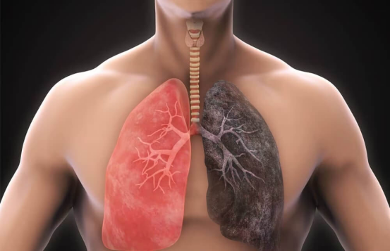
Chronic Obstructive Pulmonary Disease (COPD) remains one of the leading causes of morbidity and mortality worldwide. The progression of the disease is often punctuated by acute exacerbations (AECOPD), which are sudden worsening of respiratory symptoms that require additional therapy. Understanding the intricate link between these flares and respiratory infections is paramount for effective patient management and reducing the long-term burden on the respiratory system.
The Pathophysiology of COPD Exacerbations and Infection Risk
At the core of an exacerbation is an increase in airway inflammation, mucus production, and gas trapping. While environmental factors like pollution can trigger these events, respiratory infections—both viral and bacterial—are responsible for approximately 70% to 80% of exacerbations.
The Viral-Bacterial Synergy
Viruses such as rhinovirus, influenza, and respiratory syncytial virus (RSV) often act as the primary catalyst. These pathogens damage the epithelial lining of the lungs, impairing the mucociliary escalator. This structural damage creates a fertile environment for secondary bacterial colonization by organisms like Streptococcus pneumoniae or Haemophilus influenzae, leading to a more severe and prolonged inflammatory response.
Pro Tips: Expert Insight on Inflammation
- Monitor Sputum Color: A change from clear to purulent (yellow or green) sputum is a strong indicator of bacterial involvement and may necessitate antibiotic intervention.
- Baseline Awareness: Encourage patients to keep a daily “breathlessness diary” to distinguish between daily fluctuations and a true clinical exacerbation.

Identifying Clinical Indicators and Overlapping Symptoms
Differentiating between a standard COPD flare and one complicated by an active respiratory infection is critical for determining the treatment pathway. The “Anthonisen Criteria” remains a gold standard for assessing the severity based on three primary symptoms: increased dyspnea, increased sputum volume, and increased sputum purulence.
Diagnostic Checklist for Clinicians
- Physical examination for wheezing, tachypnea, and use of accessory muscles.
- Pulse oximetry to detect hypoxemia (Oxygen saturation below 90%).
- Chest X-rays to rule out pneumonia or pneumothorax.
- C-reactive protein (CRP) blood tests to gauge systemic inflammation levels.
| Feature | Viral Trigger | Bacterial Trigger |
|---|---|---|
| Sputum Appearance | Usually Mucoid (Clear/White) | Purulent (Yellow/Green/Thick) |
| Systemic Symptoms | High fever, muscle aches, sore throat | Localised chest congestion, productive cough |
| CRP Levels | Mildly Elevated | Significantly Elevated |
| Primary Treatment | Antivirals/Supportive Care | Antibiotics |
Comprehensive Management and Therapeutic Strategies
The management of AECOPD focuses on minimizing the impact of the current event and preventing future occurrences. This requires a multi-faceted approach involving pharmacological and supportive therapies.
Pharmacological Interventions
Short-acting beta-agonists (SABA) and short-acting muscarinic antagonists (SAMA) are the first line of defense for immediate bronchodilation. Systemic corticosteroids are often prescribed to reduce airway inflammation and improve lung function (FEV1) more rapidly. When bacterial infection is suspected, a 5-to-7-day course of antibiotics is standardized.
Oxygen Therapy and Ventilation
For patients experiencing acute respiratory failure, controlled oxygen therapy is vital. The goal is to maintain oxygen saturation between 88% and 92% to avoid hypercapnia (excess CO2). In severe cases, Non-Invasive Ventilation (NIV) is preferred over intubation as it reduces the risk of ventilator-associated pneumonia and shortens hospital stays.
Preventive Measures and Long-term Respiratory Health
Preventing the next exacerbation is as important as treating the current one. Every exacerbation causes a permanent decline in lung function, making the patient more susceptible to future infections.
Vaccination and Immunization
The annual influenza vaccine and the pneumococcal vaccine are non-negotiable components of COPD care. Recent guidelines also strongly recommend the RSV vaccine for older adults with chronic lung conditions and the Tdap vaccine to protect against pertussis.
Pulmonary Rehabilitation
Post-exacerbation pulmonary rehabilitation is one of the most effective ways to reduce hospital readmissions. It combines physical exercise, nutritional counseling, and breathing techniques to strengthen the respiratory muscles and improve the patient’s overall quality of life.
Frequently Asked Questions (FAQ)
- How do I know if my COPD flare-up is an emergency?
- Seek emergency care if you experience severe shortness of breath that does not respond to rescue inhalers, chest pain, bluish tint to lips or fingernails (cyanosis), or confusion and extreme drowsiness.
- Can a common cold lead to a COPD hospitalisation?
- Yes. In COPD patients, a viral cold can trigger significant airway inflammation, leading to an exacerbation that may require hospital treatment, especially if a secondary bacterial infection develops.
- Why are antibiotics not always prescribed for COPD flares?
- Antibiotics are only effective against bacteria. If the exacerbation is caused by a virus or environmental triggers like smoke or dust, antibiotics will not help and may cause unnecessary side effects or contribute to antibiotic resistance.
- What is the role of “Action Plans” in COPD management?
- A COPD Action Plan is a written document from your doctor that tells you exactly what to do when symptoms worsen, including when to increase inhaler use, when to start a standby prescription of steroids or antibiotics, and when to call for help.
- How long does it take to recover from an exacerbation?
- While acute symptoms may improve within 7 to 10 days, it can take several weeks or even months for a patient’s lung function and energy levels to return to their baseline state.