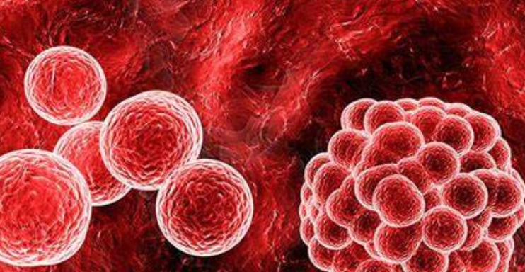
Hematopoietic Stem Cells: The Body’s Regenerative Powerhouses
At the core of our blood and immune systems lie hematopoietic stem cells (HSCs), remarkable “super cells” primarily residing within the bone marrow. These pluripotent cells are the progenitors of all blood cell types, including red blood cells, white blood cells, and platelets. Their unique capabilities of self-renewal and differentiation make them indispensable for maintaining the body’s physiological balance and responding to injury or infection. Understanding HSCs is fundamental to grasping how our body continuously replenishes its vital blood components.
The Uniqueness of Hematopoietic Stem Cells
HSCs possess two defining characteristics that set them apart from other cell types:
- Self-Renewal: This property allows HSCs to divide and produce more HSCs, ensuring a continuous supply of these foundational cells throughout a person’s life. This process is tightly regulated to prevent exhaustion or uncontrolled proliferation.
- Multi-lineage Differentiation: HSCs can mature into any type of blood cell. This differentiation is a highly complex, multi-step process influenced by various growth factors and cytokines, leading to the formation of distinct lineages: myeloid and lymphoid.
The bone marrow microenvironment, also known as the stem cell niche, plays a critical role in regulating HSC activity, providing the necessary signals for their maintenance, proliferation, and differentiation.
The Process of Hematopoiesis
Hematopoiesis is the continuous process of blood cell formation, originating from HSCs. This intricate biological pathway ensures that the body always has an adequate supply of functional blood cells. The process can be broadly divided into several stages:
- Common Myeloid Progenitor (CMP): HSCs first differentiate into CMPs, which then give rise to red blood cells (erythrocytes), platelets (thrombocytes), granulocytes (neutrophils, eosinophils, basophils), and monocytes.
- Common Lymphoid Progenitor (CLP): HSCs also differentiate into CLPs, which are the precursors for lymphocytes, specifically T cells, B cells, and Natural Killer (NK) cells, crucial components of the adaptive immune system.
Each step in this differentiation pathway is precisely controlled, involving specific gene expression patterns and cellular interactions to ensure the correct cell types are produced in appropriate numbers.
Clinical Significance and Therapeutic Applications
The profound capabilities of HSCs have made them a cornerstone in medical treatments, particularly in the field of transplantation. Hematopoietic stem cell transplantation (HSCT), often referred to as bone marrow transplantation, is a life-saving procedure for patients with various hematological malignancies, severe aplastic anemia, and certain immune deficiencies. In this procedure, healthy HSCs are infused into a patient whose own bone marrow has been compromised or destroyed, allowing for the reconstitution of a healthy blood and immune system. Research continues to explore the potential of HSCs in regenerative medicine, aiming to harness their regenerative properties for broader therapeutic applications.

Red Blood Cells: The Essential Oxygen Carriers
Among the most abundant cells in the human body, red blood cells (erythrocytes) are the primary vehicles for oxygen transport from the lungs to every tissue and organ. Their unique biconcave disc shape and lack of a nucleus in mature form are adaptations that maximize their efficiency in carrying oxygen and navigating through the body’s smallest capillaries. Without the tireless work of red blood cells, cellular respiration—the process that powers all bodily functions—would cease.
Structure and Function of Erythrocytes
The distinct morphology of red blood cells is directly linked to their critical function:
- Biconcave Shape: This shape provides a large surface area-to-volume ratio, facilitating rapid gas exchange. It also grants flexibility, allowing red blood cells to deform and squeeze through narrow capillaries, some of which are smaller than the cell itself.
- Hemoglobin Content: The cytoplasm of red blood cells is packed with hemoglobin, an iron-rich protein that binds reversibly to oxygen and carbon dioxide. Each hemoglobin molecule can carry four oxygen molecules, giving blood its characteristic red color when oxygenated.
- Anucleated: Mature red blood cells eject their nucleus and most organelles during maturation. This frees up space for more hemoglobin and prevents the cell from consuming the oxygen it is meant to transport. However, it also means they cannot repair themselves, leading to a finite lifespan.
The primary function of red blood cells is to deliver oxygen from the pulmonary capillaries to systemic tissues and to transport carbon dioxide, a waste product of metabolism, back to the lungs for exhalation. This gas exchange is vital for maintaining cellular energy production and acid-base balance.
Erythropoiesis: The Formation of Red Blood Cells
The production of red blood cells, known as erythropoiesis, is a carefully regulated process that primarily occurs in the bone marrow in adults. It is a continuous cycle, ensuring that old or damaged red blood cells are replaced efficiently. The key steps include:
- HSC to Erythroid Progenitor: Hematopoietic stem cells differentiate into erythroid progenitor cells under the influence of various growth factors.
- Proerythroblast to Reticulocyte: These progenitors develop through several stages (proerythroblast, basophilic erythroblast, polychromatophilic erythroblast, orthochromatophilic erythroblast), gradually synthesizing hemoglobin and decreasing in size.
- Nucleus Ejection and Reticulocyte Release: In the final stages, the nucleus is ejected, and the cell becomes a reticulocyte, which is then released into the bloodstream. Reticulocytes mature into full erythrocytes within 1-2 days.
The hormone erythropoietin (EPO), primarily produced by the kidneys, is the main regulator of erythropoiesis. Low oxygen levels (hypoxia) stimulate EPO release, which in turn boosts red blood cell production.
Red Blood Cell Lifecycle and Breakdown
The average lifespan of a red blood cell is approximately 120 days. As they age, their membranes become less flexible, and they are eventually recognized and removed from circulation by macrophages, primarily in the spleen and liver. This breakdown process involves:
- Hemoglobin Recycling: Hemoglobin is broken down into heme and globin. Globin is further broken into amino acids, which are reused. Heme is converted into biliverdin, then bilirubin, which is transported to the liver and excreted in bile.
- Iron Storage and Reuse: The iron released from heme is bound to transferrin and transported to the bone marrow for reuse in new hemoglobin synthesis or stored as ferritin in the liver.
Efficient recycling mechanisms ensure that valuable components are not wasted, highlighting the body’s remarkable ability to conserve resources.

Monocytes & Immune Cells: The Body’s Defense System
The human body is under constant threat from pathogens, toxins, and abnormal cells. Protecting us from these dangers is a sophisticated and multi-layered defense system known as the immune system, comprised of various specialized cells. Among these, monocytes play a crucial role as frontline defenders, acting as versatile phagocytes and precursors to other important immune cells. Understanding the intricate workings of monocytes and the broader immune cell landscape is key to appreciating how our body maintains its integrity and health.
Monocytes: The “Big Guys” of the Blood
Monocytes are the largest type of white blood cell, circulating in the bloodstream for a few days before migrating into tissues, where they differentiate into macrophages and dendritic cells. This transformation significantly enhances their defensive capabilities:
- Phagocytosis: Monocytes, and especially their macrophage derivatives, are highly effective phagocytes. They engulf and digest foreign particles, cellular debris, and pathogens like bacteria and viruses. This process is essential for clearing infections and maintaining tissue homeostasis.
- Antigen Presentation: Macrophages and dendritic cells are professional antigen-presenting cells (APCs). They process ingested pathogens and present fragments (antigens) on their surface to T lymphocytes, thereby initiating and shaping adaptive immune responses.
- Cytokine Production: Monocytes and macrophages release various cytokines, signaling molecules that regulate inflammation, recruit other immune cells to the site of infection, and influence immune cell differentiation and activity.
Their ability to patrol tissues and adapt their function makes monocytes critical components of both innate and adaptive immunity, bridging the gap between the body’s immediate defense and its targeted, long-term responses.
The Broader Spectrum of Immune Cells
The immune system involves a diverse array of cells, each with specialized functions:
| Immune Cell Type | Primary Function | Role in Defense |
|---|---|---|
| Neutrophils | First responders to infection | Phagocytosis of bacteria, release of antimicrobial substances |
| Eosinophils | Combat parasitic infections, allergic reactions | Release toxic granules to kill parasites, modulate allergic responses |
| Basophils/Mast Cells | Allergic and inflammatory responses | Release histamine and other mediators |
| Lymphocytes (T cells, B cells, NK cells) | Adaptive immunity, viral defense | T cells: direct killing of infected cells, regulate immune response. B cells: produce antibodies. NK cells: kill tumor cells and virus-infected cells. |
| Macrophages | Phagocytosis, antigen presentation | Clear pathogens, present antigens to T cells, tissue repair |
| Dendritic Cells | Most potent antigen-presenting cells | Initiate primary immune responses by activating T cells |
This cellular orchestra works in concert to identify, neutralize, and remember threats, providing both immediate protection and long-lasting immunity.
How the Body Defends Itself Against Pathogens
The body’s defense against invading pathogens involves a coordinated effort between innate and adaptive immunity:
- Innate Immunity (Non-specific): This is the body’s first line of defense, providing immediate, broad-spectrum protection. It includes physical barriers (skin, mucous membranes), chemical barriers (stomach acid, tears), and cellular components like neutrophils, macrophages, and NK cells. These cells recognize common pathogen-associated molecular patterns (PAMPs) and rapidly eliminate threats.
- Adaptive Immunity (Specific): This highly specific defense system develops over time and targets particular pathogens. It involves lymphocytes (T and B cells) that recognize specific antigens. Upon first exposure, it mounts a primary response, and upon subsequent exposure, it generates a faster and stronger secondary response, leading to immunological memory.
The interplay between these two systems ensures a robust and adaptable defense, capable of protecting us from a vast array of microbial threats and maintaining our health.

Liver Cells (Hepatocytes): Metabolic Powerhouses & Detoxifiers
The liver, a vital organ weighing about 3 pounds, is a true multi-tasker, performing over 500 essential functions that are critical for survival. At the heart of these functions are liver cells, or hepatocytes, which constitute approximately 80% of the liver’s mass. These highly specialized cells are responsible for an incredible array of metabolic, synthetic, and detoxifying processes, effectively acting as the body’s central processing unit for nutrients, waste products, and foreign substances. Understanding their complex roles reveals how the liver protects and maintains overall health.
Diverse Metabolic Functions of Hepatocytes
Hepatocytes are unequaled in their metabolic versatility, playing pivotal roles in the metabolism of carbohydrates, fats, and proteins:
- Carbohydrate Metabolism: The liver is central to maintaining stable blood glucose levels.
- Glycogenesis: Converts excess glucose into glycogen for storage.
- Glycogenolysis: Breaks down stored glycogen into glucose when blood sugar is low.
- Gluconeogenesis: Synthesizes new glucose from non-carbohydrate sources (e.g., amino acids, glycerol) during prolonged fasting.
- Lipid Metabolism: Hepatocytes synthesize cholesterol, lipoproteins (HDL, LDL, VLDL), and bile acids. They also break down fatty acids to produce energy and convert excess carbohydrates and proteins into fatty acids for storage.
- Protein Metabolism: The liver is the primary site for the synthesis of most plasma proteins, including albumin (maintains osmotic pressure), clotting factors, and transport proteins. It also deaminates amino acids, converting ammonia (a toxic byproduct) into urea for excretion (the urea cycle).
These intricate metabolic pathways ensure that the body has a steady supply of energy, building blocks, and regulatory molecules, while also managing waste products.
Detoxification and Biotransformation
One of the liver’s most critical functions is to neutralize and eliminate harmful substances from the body. Hepatocytes are equipped with extensive enzymatic machinery, particularly in their smooth endoplasmic reticulum and peroxisomes, to perform detoxification and biotransformation:
- Phase I Reactions (Functionalization): Involve enzymes like cytochrome P450 (CYP) that add or expose polar groups on lipophilic compounds (drugs, toxins, hormones), making them more reactive for subsequent reactions.
- Phase II Reactions (Conjugation): Involve enzymes that conjugate (attach) polar molecules (e.g., glucuronic acid, sulfate, glutathione) to the modified compounds, significantly increasing their water solubility. This makes them easier to excrete via bile or urine.
This two-phase process effectively converts toxic, lipid-soluble substances into non-toxic, water-soluble forms that can be safely removed from the body. This includes drugs, alcohol, environmental toxins, and metabolic waste products.
Bile Production and Digestive Support
Hepatocytes also produce bile, a greenish-yellow fluid essential for digestion and absorption of fats in the small intestine. Bile contains bile salts, cholesterol, bilirubin, and phospholipids. Bile salts emulsify dietary fats, breaking them down into smaller droplets, which increases the surface area for lipase enzymes to act upon. This facilitates fat digestion and the absorption of fat-soluble vitamins (A, D, E, K). Additionally, bile serves as a route for the excretion of bilirubin and other waste products.
Impact on Overall Health
The health of hepatocytes directly reflects the overall health of an individual. Liver dysfunction, caused by factors such as viral infections (hepatitis), alcohol abuse, fatty liver disease, or drug toxicity, can severely impair these vital functions, leading to systemic metabolic disturbances, accumulation of toxins, and ultimately, life-threatening conditions. Maintaining liver health through a balanced diet, limiting alcohol intake, and avoiding hepatotoxic substances is paramount for long-term well-being.

Neurons & Muscle Cells: The Architects of Communication and Movement
The ability to think, perceive, move, and react fundamentally relies on the specialized functions of neurons (nerve cells) and muscle cells (myocytes). These two cell types work in a sophisticated partnership, with neurons transmitting electrical signals that command muscle cells to contract, thereby enabling all forms of bodily movement and internal organ function. Exploring the intricate mechanisms of these cells unveils the biological basis of consciousness, sensation, and physical activity.
Neurons: The Body’s Electrical Messengers
Neurons are the fundamental units of the nervous system, responsible for transmitting information throughout the body via electrical and chemical signals. Their unique structure facilitates this rapid communication:
- Cell Body (Soma): Contains the nucleus and most cellular organelles, responsible for the neuron’s metabolic activities.
- Dendrites: Branch-like extensions that receive incoming signals from other neurons or sensory receptors. They increase the surface area for synaptic connections.
- Axon: A long, slender projection that transmits electrical signals (action potentials) away from the cell body to other neurons, muscles, or glands. Axons can be myelinated, meaning they are covered by a fatty sheath that insulates the axon and speeds up signal transmission.
- Axon Terminals (Synaptic Buttons): Endings of the axon that form synapses with other cells, releasing neurotransmitters to transmit signals across the synaptic cleft.
The primary mechanism of neuronal communication is the action potential, a brief, rapid change in the electrical potential across the neuron’s membrane. This “all-or-none” electrical impulse travels down the axon to the synapse, where it triggers the release of neurotransmitters. These chemical messengers bind to receptors on the target cell, either exciting or inhibiting its activity. This electrochemical signaling forms the basis of all nervous system functions, from simple reflexes to complex cognitive processes.
Muscle Cells: The Engines of Contraction
Muscle cells, or myocytes, are specialized for contraction, generating force and movement. There are three main types of muscle cells, each with distinct characteristics and functions:
- Skeletal Muscle Cells: These are long, cylindrical, multi-nucleated cells that are responsible for voluntary movements. They are organized into bundles and attached to bones via tendons. Their contraction is initiated by signals from motor neurons.
- Cardiac Muscle Cells: Found exclusively in the heart, these cells are branched, typically uni-nucleated, and interconnected by intercalated discs that allow for synchronized contraction. Cardiac muscle contraction is involuntary and intrinsic, though regulated by the nervous system.
- Smooth Muscle Cells: These are spindle-shaped, uni-nucleated cells found in the walls of internal organs (e.g., digestive tract, blood vessels, bladder). Their contraction is involuntary and responsible for processes like peristalsis, blood pressure regulation, and pupil dilation.
The fundamental mechanism of muscle contraction involves the interaction of two main proteins: actin and myosin. In skeletal and cardiac muscle, these proteins are arranged into repeating units called sarcomeres, giving the muscle a striated appearance. When a muscle cell receives an electrical signal (often from a neuron), calcium ions are released, triggering the myosin heads to bind to actin filaments and pull them past each other, shortening the sarcomere and thus the muscle cell. This intricate sliding filament model is powered by ATP.
The Neuromuscular Junction: Bridging Communication and Contraction
The crucial link between the nervous system and the muscular system occurs at the neuromuscular junction. Here, the axon terminal of a motor neuron forms a specialized synapse with a muscle fiber. When an action potential reaches the axon terminal, it causes the release of the neurotransmitter acetylcholine. Acetylcholine binds to receptors on the muscle cell membrane, triggering another action potential in the muscle cell, which then propagates and leads to muscle contraction. This precise and rapid communication ensures that neural commands are efficiently translated into physical actions, allowing for everything from intricate finger movements to powerful leaps.

Cellular Interconnectedness: A Symphony of Life
While we have explored various cell types individually, it is crucial to recognize that no cell in the human body operates in isolation. From the initial differentiation of hematopoietic stem cells to the coordinated contractions driven by neurons and muscle cells, all cellular functions are intricately linked and interdependent. This profound interconnectedness forms a complex biological symphony, where each cell type plays a vital role in maintaining the overall harmony and health of the organism. Understanding this cellular dialogue is paramount to comprehending human physiology and pathology.
The Dynamic Balance of Cellular Systems
The body maintains a delicate balance, or homeostasis, through constant communication and regulation among different cell types and organ systems. Consider the following examples:
- Oxygen Transport and Metabolism: Red blood cells transport oxygen, which is essential for liver cells to perform their vast metabolic and detoxification functions. Neurons and muscle cells also heavily rely on a steady oxygen supply to generate ATP for their electrical signaling and contractile activities. Without efficient oxygen delivery, all these systems would fail.
- Immune Surveillance and Tissue Repair: When an infection occurs, immune cells, including monocytes and their macrophage derivatives, not only fight pathogens but also communicate with other cells to initiate tissue repair. HSCs are crucial for replenishing immune cells, ensuring a continuous defense capability.
- Nutrient Distribution and Waste Removal: Liver cells process nutrients absorbed from the digestive system, making them available to all other cells. They also detoxify waste products, which are then excreted, often with the help of the kidneys, ensuring a clean internal environment for optimal cellular function.
This constant exchange of nutrients, signals, and waste products underscores the deep interdependencies that exist at the cellular level, ensuring that the body functions as a unified, coherent whole.
Cellular Communication Pathways
Cells communicate through a variety of mechanisms, including:
- Direct Contact: Cells can communicate directly through gap junctions (in animal cells) or plasmodesmata (in plant cells), allowing for the direct passage of small molecules and ions. Immune cells also make direct contact with target cells or antigen-presenting cells.
- Chemical Signaling (Paracrine, Endocrine, Autocrine):
- Paracrine: Cells release signaling molecules that act on nearby cells.
- Endocrine: Cells release hormones into the bloodstream, which travel to distant target cells (e.g., hormones regulating cellular metabolism).
- Autocrine: Cells release signals that act back on themselves, often involved in regulating cell growth and differentiation.
- Synaptic Signaling: Specialized communication between neurons and their target cells (other neurons, muscle cells, glands) via neurotransmitters at synapses, enabling rapid and precise information transfer.
These diverse communication pathways ensure that cells can coordinate their activities, respond to environmental changes, and maintain the complex processes necessary for life.
The Unifying Principle of Cellular Life
Ultimately, the human body is a testament to the power of cellular specialization and cooperation. Each cell, no matter how unique its structure or function, contributes to the larger goal of sustaining life. From the microscopic scale of molecular interactions within a single hepatocyte to the macroscopic scale of organ system function, the underlying principle is one of complex, emergent behavior arising from the coordinated actions of billions of individual cells. Continued research into cellular biology promises to unlock even deeper understanding of health, disease, and the very essence of human existence.
Frequently Asked Questions (FAQ)
- Q1: What is the primary role of hematopoietic stem cells
Reader safety note: This article is general educational content only. It is not medical advice, mental-health diagnosis, psychotherapy, public-health guidance, emergency help, medication guidance, device instruction, or individualized relationship advice. Consult qualified medical, mental-health, public-health, legal, educational, or safety professionals for personal decisions, urgent symptoms, self-harm risk, abuse, addiction, infectious disease concerns, or treatment questions.